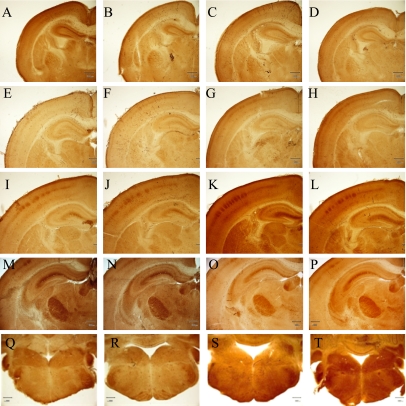
Fig. 3.
Cortical barrel, barreloid and barrelette at P1, P4 and P8. Cortical barrels at P1 (A–D), P4 (E–H) and at P8 (I–L): control males (A, E, I), control females (B, F, J), BPA-treated males (C, G, K) and BPA-treated females (D, H, L), respectively. Barreloid (M–P) and barrelette at P8 (Q–T): control males (M, Q), control females (N, R), BPA-treated males (O, S) and BPA-treated females (P, T), respectively. COX staining. Bar=300 µm.

