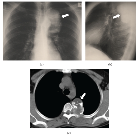
Figure 1.
A 32-year-old man with chondrosarcoma. The posteroanterior (a) and lateral (b) chest radiographs, show a well-defined radiopaque lesion in the left posterior paraspinal location (arrows). (c) The axial MDCT image demonstrates a soft-tissue mass (arrow) with amorphous “rings and arcs” calcified matrix (thin arrow) and adjacent neural foramina widening (asterisk).

