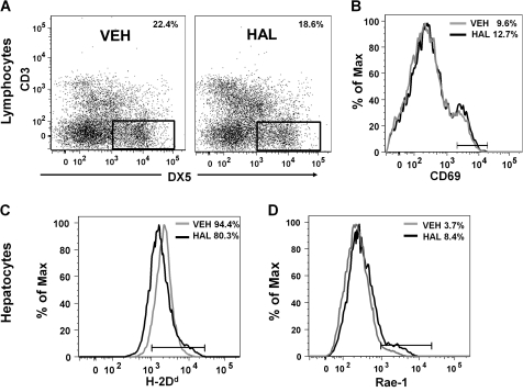
FIG. 6.
HAL treatment alters the phenotype of hepatic NK cells and hepatocytes in vivo. (A) Hepatic lymphocytes from VEH- or HAL-treated mice were pooled according to their respective groups. NK cells were gated as DX5+, CD3− as indicated in the dot plot graphs. The percentage of NK cells in the population is noted in each plot. Similar results were obtained in three independent experiments. (B) NK cell surface expression of CD69 was analyzed by flow cytometry. The gray and black histograms depict the VEH and HAL treatment groups, respectively. Experimental replicate had similar results. Hepatocytes isolated from VEH- and HAL-treated mice were immunolabeled with anti-H2Dd (C) and anti-Rae-1 (D) antibodies and analyzed by flow cytometry. The gray and black histograms depict the VEH and HAL treatment groups, respectively. Similar results were obtained in four independent replicates.

