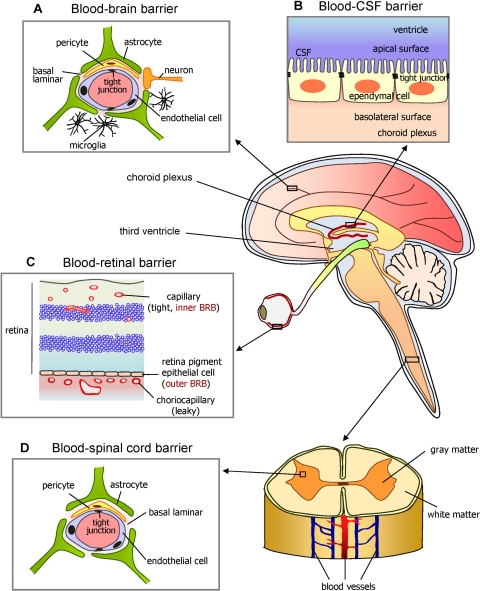
Figure 1.
Diagram of the brain and spinal cord illustrating how the eye's inner and outer blood-retinal barriers (BRBs) fit into the overall scheme of blood-neural barriers (BNBs). Barriers between the blood and neural tissues are collectively referred to as BNBs and include the blood-brain barrier (A), the blood-CSF barrier (B), the BRB (C), and the blood-spinal cord barrier (D). Other similar specialized barriers in the body include the blood-labyrinth barrier in the inner ear, the blood-nerve barrier in the peripheral nervous system, and the blood-testis barrier. Their function is to act as semipermeable paracellular passive diffusion barriers or gates to large and hydrophilic solutes. Small, lipophilic essential nutrients and toxic metabolites are delivered or removed, respectively, through passive or active site-specific transcellular carrier–mediated influx or efflux transporters. Considering that several retinal disorders are accompanied by dysfunction or breakdown of this BRB and their associated cell-cell signaling mechanisms, elucidating the nature of the BRB is important for understanding normal health and disease. The static morphologic structure responsible for all these organ-specific barriers is the tight junction (TJ). Counterintuitively, curative drug therapy to these protected sites requires that drugs circumvent these naturally protective barriers. Reprinted with permission from Choi YK, Kim KW. Blood-neural barrier: its diversity and coordinated cell-to-cell communication. BMB Rep. 2008;41:345–352.

