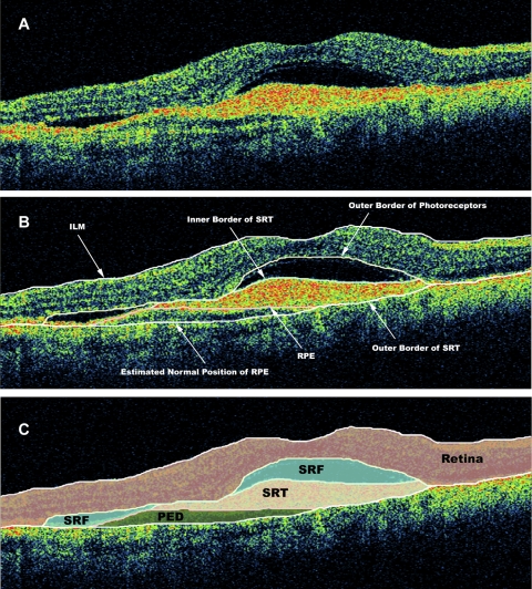
Figure 1.
OCT B-scan demonstrating (A) subretinal tissue, SRF, and PED. (B) The clinically relevant boundaries—the internal limiting membrane (ILM), the outer border of the photoreceptors, the RPE, the inner border of the subretinal tissue, the outer border of the subretinal tissue (SRT), and the estimated normal location of the RPE layer—were graded by OCTOR (computer-assisted manual grading) software. (C) The software then computed the volumes of the spaces (retina, SRT, SRF, and PED) defined by these boundaries.

