Figure 8.
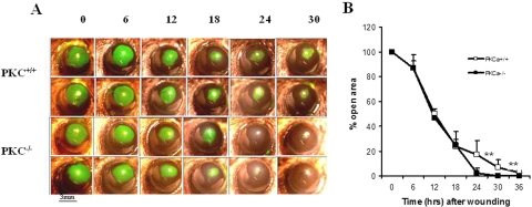
Accelerated corneal epithelial wound healing in PKCα−/− mice. (A) Fluorescein-stained corneal images after corneal epithelial debridement in PKCα+/+ and PKCα−/− mice. (B) Percentage of the original wound area is plotted over time and was calculated from the area of fluorescein staining at each time point. Data are mean ± SD (n = 6, **P < 0.01).

