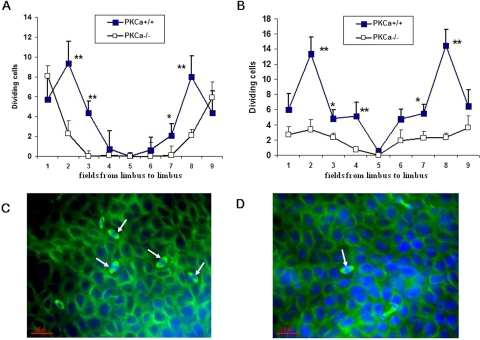
Figure 9.
Effects of PKCα deficiency on dividing basal cells after corneal epithelial debridement. Dividing basal epithelial cells in each region of the cornea at 18 (A) and 30 (B) hours, respectively, after epithelial debridement. Representative photos of dividing cells (arrow) stained by tubulin (green) in PKCα+/+ (C) and PKCα−/− mice (D) at 18 hours after wounding. DAPI (blue) was used for counterstaining. *P < 0.05 and **P < 0.01.

