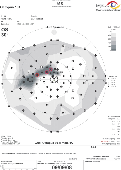
Figure 3.
Threshold-estimating static perimetry with regional stimulus condensation in the superior paracentral visual field clearly demarcates a circumscribed paracentral small retinal nerve fiber-related scotoma. Circles: 30-A grid with polar test point arrangement. Only three pathologic locations were detected without condensed stimulus arrangement (red circles).

