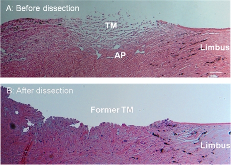
Figure 2.
Hematoxylin and eosin–stained paraffin-embedded sagittal sections of the angle region from porcine eyes. (A) Sample with the iris removed and with the corneal endothelium and iris root scraped but with the trabecular meshwork and aqueous plexus region untouched. (B) Sample after harvesting the trabecular meshwork and aqueous plexus tissue for cell culture, showing tissues that were cut away during dissection. Scale bar, 100 μm.

