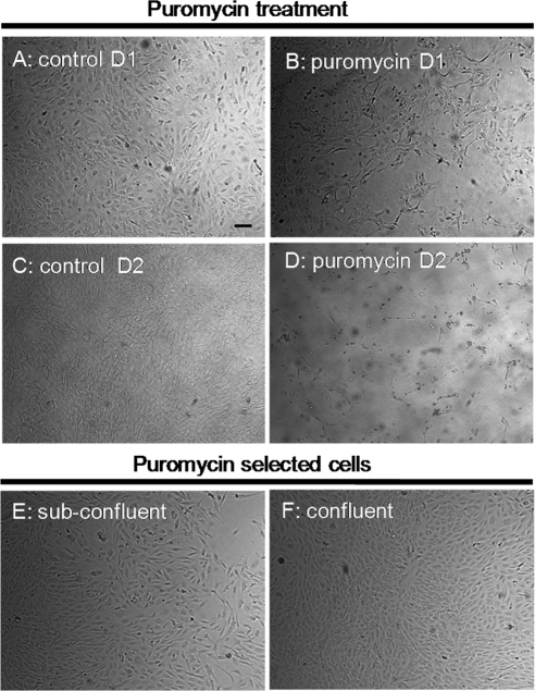
Figure 4.
Phase-contrast microscopy of porcine cells from outflow tissues after exposure to puromycin. Approximately two-thirds of the cells died on treatment with puromycin (B) for 1 day (D1) compared with control (A). After 2 days (D2) of exposure, the number of dead cells increased significantly (D) compared with control cells (C). After the selection process, the remaining cells appeared healthy, both at 6 days (E) and at 10 days (F) after the removal of puromycin. At confluence, the selected cells displayed a cobblestone appearance (F). Shown are images from 1 of 6 experiments. All images are shown at the same magnification. Scale bar, 50 μm.

