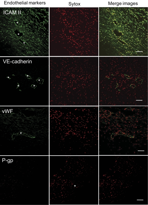
Figure 7.
Expression of endothelial marker proteins in confocal sections through porcine aqueous plexus region. Confocal immunofluorescence microscopy shows expression of ICAM II, VE-cadherin, and vWF by aqueous plexus lining cells. Asterisks: AP lumen. We were unable to detect P-gp expression using this methodology. Endothelial marker protein (top left of each image) is shown in green, and nuclei are shown in red. Data are representative images from three porcine eyes analyzed. Scale bars, 50 μm.

