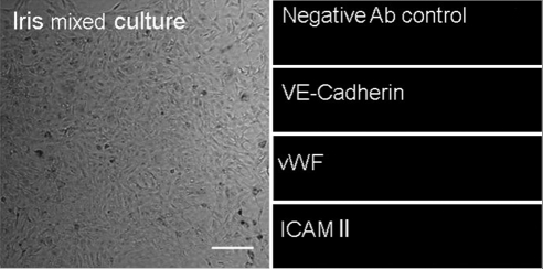
Figure 8.
Phase-contrast and confocal microscopy of the isolated iris cells that were not exposed to puromycin. Iris cells appear more pigmented and display a mixture of morphologic features (left). Moreover, we were unable to detect any of the endothelial markers expressed by AAP cells (i.e., vWF). Shown are representative images of 1 of 3 experiments. All images are shown at the same magnification. Scale bar, 100 μm.

