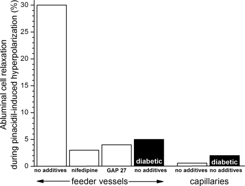
Figure 9.
Percentage of monitored abluminal cells that relaxed during hyperpolarization induced by 5 μM pinacidil, an activator of microvascular KATP channels.5,38 For the nifedipine-containing group, microvessels were pre-exposed to solution A supplemented with 10 μM nifedipine for 10 to 15 minutes. For the GAP 27 group, microvessels were pre-exposed to solution A supplemented with 300 μM GAP 27 for 2 hours; 60- to 90-minute exposure to this concentration of GAP 27 eliminated gap junction transmission in retinal microvessels.17 For feeder vessels, the number of monitored abluminal (mural) cells was 66, 29, 53, and 166 for the no additives, nifedipine, GAP 27, and diabetic/no additives group, respectively. For capillaries, the number of monitored abluminal cells (pericytes) was 25 and 49 for the no additives and the diabetic/no additives group, respectively.

