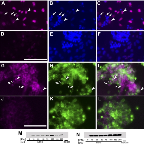
Figure 2.
BrdU labeling and TAg expression in conditionally immortalized retinal cells. (A–F) BrdU immunostaining of primary, mixed retinal cultures in growth medium with 50 U/mL IFNγ at 33°C showing (A, D) BrdU, (B, E) Hoechst, and (C, F) overlay. (A–C) BrdU (10 μM) added 17 hours before fixation and immunostaining. Large arrowheads: large BrdU-positive nuclei with typical diffuse Hoechst staining. Arrows: small BrdU-negative nuclei with uniform and intense Hoechst staining. (D–F) Control wells grown in parallel without BrdU. (G–L) Photomicrographs of mixed retinal cultures showing (G, J) immunostaining for SV40-TAg and (H, K) green fluorescence protein (HRhoGFP) expression. (I, L) Overlay. (G–I) Cells cultured with 50 U/mL IFNγ at 33°C for 4 days. Arrows: TAg-positive, HRhoGFP-negative (nonphotoreceptor) cells. Large arrowheads: TAg-positive, HRhoGFP-positive (rod photoreceptors) cells. Small arrowheads: TAg-negative, HRhoGFP-positive cells. (J–L) Cells cultured with 0 U/mL IFNγ at 39°C for 4 days. Scale bars, 50 μm. (M) Western blot analysis of TAg expression in ImM10 cells cultured in growth medium containing 0 to 200 U/mL IFNγ for 4 days in the presence of 0 to 200 U/mL IFNγ and in HEK293 cells (negative control). (N) Longer exposure of blot shown in (A) reveals trace levels of TAg expression in ImM10 cells cultured without IFNγ but none in HEK293. Increased intensity of TAg expression at 100 U/mL reflects increased loading of total proteins in that lane.

