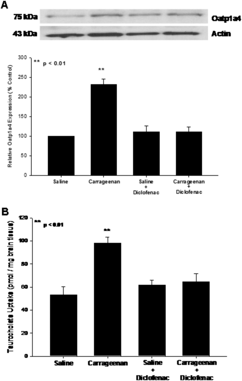
Fig. 6.
Effect of diclofenac, a nonsteroidal anti-inflammatory drug, on Oatp1a4 functional expression. A, Western blot analysis of microvessels isolated from rats treated with saline or λ-carrageenan in the presence and absence of diclofenac. Crude membrane preparations from rat brain microvessels (10 μg) were resolved on a 10% lithium dodecyl sulfate-polyacrylamide gel and transferred to a PVDF membrane. Samples were analyzed for expression of Oatp1a4 using the polyclonal antibody M-50 (1:500 dilution). Relative levels of Oatp1a4 were determined by densitometric analysis. Results are expressed as mean ± S.D. of three separate experiments. Asterisks represent data points that are significantly different from control. B, uptake of taurocholate into rat brain after 3-h λ-carrageenan-induced inflammatory pain in the presence and absence of diclofenac. Graph shows the concentration of taurocholate detected in rat brain tissue for the four treatment groups after injection of 3% λ-carrageenan or 0.9% saline into the plantar surface of the right hind paw. Diclofenac (30 mg/kg) was injected 15 min after footpad injection. Results are expressed as mean ± S.D. of six animals per treatment group. Asterisks represent data points that are significantly different from control.

