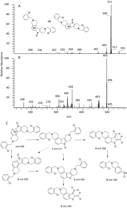
Fig. 3.
Mass spectra of TSOD (M6) in a positive ion mode. A, MS/MS spectrum of m/z 559 ion (MH+ containing 35Cl isotope. B, mass spectrum of m/z 559 ion (MS3) obtained in a data-dependent mode after fragmentation of m/z 511 ion. C, structures of fragment ions observed in the MS/MS and MS3 spectra of m/z 559. The proposed structure of only one isomer is shown. The positional isomers of TSOD could not be differentiated from the mass spectral fragment ions.

