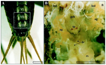
Figure 1.
A) Terminal abdominal segments (ventral view) of female T. commodus. Segments containing the genital chamber and associated glands of the reproductive system are marked by Roman numbers, while the dashed frame indicates the position of the organs exhibited in image B. B) In-vivo position of selected reproductive organs in T. commodus. Abbreviations: ag, accessory glands; bl, basal lamina; cb, cell border; cfc, cuticula-forming cell; ci, cuticular intima; cod, common oviduct; dm, desmosome; dr, ductus receptaculi; dr2, ductus receptaculi, Region III; dr3, ductus receptaculi, Region III; ecs, extracellular space; ed, efferent ductule; ep, epithelium; ft, fatty tissue; gch, genital chamber; gc, glandular cell; int, cellular interdigitations; lod, lateral oviduct; l, lumen; mc, muscle coat; mit, mitochondrium; mt, muscle tissue; mv, microvilli; n, nucleus; nl, nucleolus; op, ovipositor; rs, receptaculum seminis; ser, smooth endoplasmatic reticulum; tp, terminal papilla; ves, vesicle. Bars: l mm.

