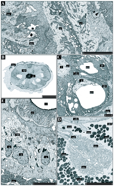
Figure 5.
Detailed illustrations of the histology and ultrastructure of the ductus receptaculi. Region I (A) is chiefly characterized by a zipper-like longitudinal section of the lumen (arrowheads) preventing an uncontrolled outflow of spermatozoa from the receptaculum. Region II (B-D) consists of glandular cells (gc) producing the ductal secretions and cuticula-forming cells (cfc) being responsible for the formation of the cuticular intima (ci). The gland cells contain an extracellular space (ecs), into which the secretions are re leased and transported towards the intima through an efferent ductule (ed). The cavity is surrounded by a dense layer of microvilli (mv). Region III (E) consists of uniformly structured, cubic to columnar epithelial cells, an inhomogeneously structured cuticular intima, and a rather wide lumen (I). Abbreviations: see Figure I legend. Bars: 30 µm in B, 5 µm in A and C-E.

