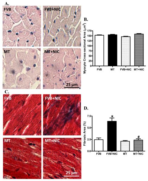
Fig. 5.
H&E and Masson trichrome stained photomicrographs exhibiting cardiomyocyte area and myocardial fibrosis, respectively, in myocardium from FVB and MT mice treated with or without nicotine (2 mg/kg/d for 10 days, i.p.). Panel A: H&E staining in FVB and MT groups with or without nicotine treatment; Panel B: Pooled data of cardiomyocyte cross-sectional area; Panel C: Masson trichrome staining exhibiting myocardial fibrosis; and Panel D: Pooled data of myocardial fibrosis. Mean ± SEM, n = 10–15 fields from three mice per group, * p < 0.05 vs. FVB group, # p < 0.05 vs. FVB+NIC group.

