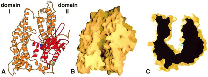
Figure 1.
Overview of the rat neurolysin structure. (A) Ribbon view of the molecule looking down on the substrate-binding channel. The active site zinc ion is shown as a blue sphere, and the region structurally similar to other metallopeptidases is shown in red. (B) The same view shown as a molecular surface representation. (C) Cross-sectional view of the neurolysin substrate-binding channel. The depth of the channel at the active site (zinc ion in blue) is approximately 35 Å. This figure and all other ribbon and surface figures were prepared with molscript (50) and grasp (51).

