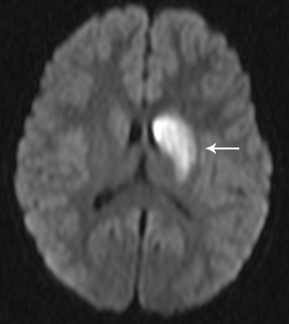
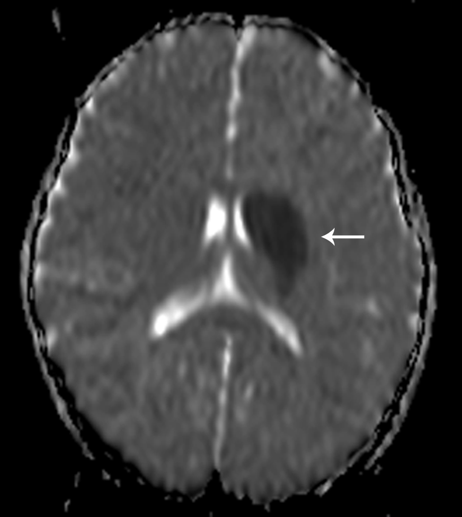
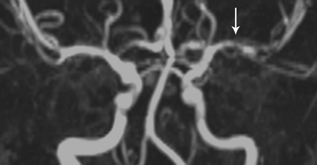
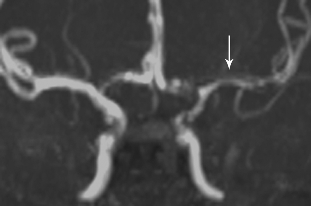
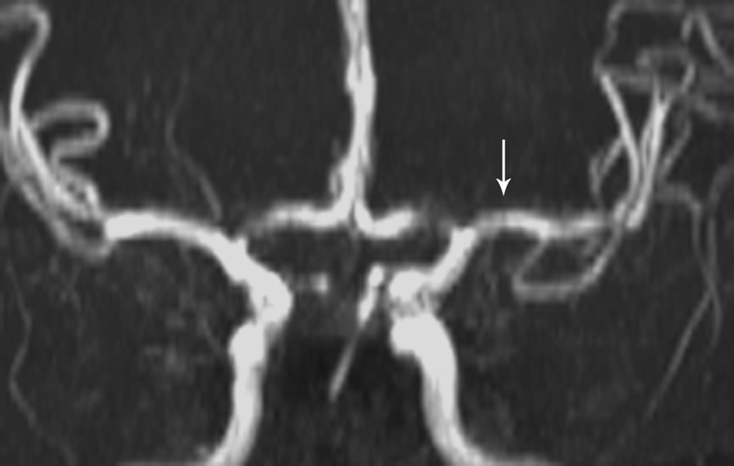
Fig 1. Focal Cerebral Arteriopathy.
8-year-old patient with MRI demonstrating acute stroke in the left basal ganglia that is bright on diffusion-weighted imaging (DWI) (Figure 1a) and dark on the apparent diffusion coefficient (ADC) map (Figure 1b). Magnetic resonance angiogram (MRA) of the circle of Willis shows a focal stenosis of the left middle cerebral artery (MCA) M1 segment (arrow) (Figure 1c). This appears worse 3 weeks later (Figure 1d) and better 1 year after stroke, though continued mild narrowing of the left MCA M1 segment is present (Figure 1e)

