
Figure 4.
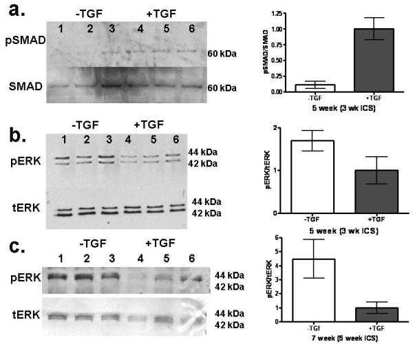
a: Western blotting for ERK 1/2 and SMAD activation of nHDF in constructs with and without TGF-β1 supplementation at 5 weeks. a: Western blot for SMAD and phosphorylated SMAD (pSMAD). Western Blot for levels of total ERK (tERK) and threonine 202/tyrosine204 phosphorylated ERK (pERK) at b: 5 weeks and c: 7 weeks. Band intensity was determined by densitometry and expressed as the ratio of phosphorylated protein band to total protein band and then normalized to the +TGF-β value. TGF-β is shown to increase fraction of SMAD that is phosphorylated but decrease the fraction of ERK that is phosphorylated. See Fig. 1 regarding paired symbols (n=3).