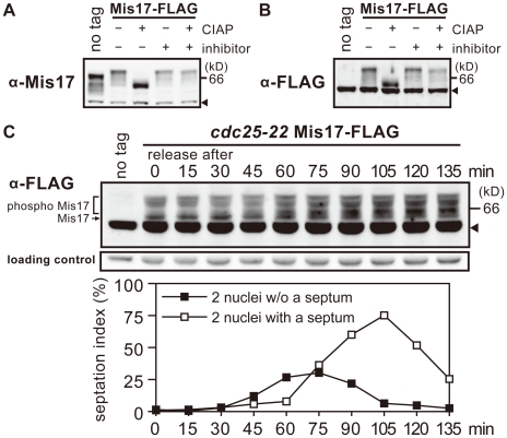
Figure 1. Mis17 is a hyperphosphoprotein.
A–B. An SDS-PAGE of S. pombe cell extracts was performed to detect Mis17 and Mis17-FLAG by immunoblot, using antibodies against bacterial-made Mis17 (α-Mis17 in A) and FLAG (α-FLAG in B). The wild-type strains with no tag and the chromosomally-integrated Mis17-FLAG gene expressed under the native promoter were used. Cell extracts of the Mis17-FLAG strain were treated with the calf intestine alkaline phosphatase (CIAP) in the presence (+) or absence (−) of phosphatase inhibitors (β-glycerophosphate and p-nitrophenyl phosphate). Arrowheads indicate the cross-reacting antigens. The no-tag band migrated faster than the Mis17-FLAG band with the expected MW difference. C. Mis17-FLAG chromosomally integrated in the cdc25-22 mutant and expressed under the native promoter was detected by immunoblot at 15 min intervals from 0–135 min (top). Arrowhead indicates the cross-reacting antigens. A shorter exposed image of the cross-reacting antigens is shown as the loading control (middle). The mutant cells were arrested in the G2 phase at 36°C, and released to mitosis by shifting the temperature to 26°C. Cells synchronously progressed with the timing of chromosome segregation, septation, and cytokinesis at 45, 75, and 105 min, respectively (bottom). Frequencies of cells showing two nuclei without (filled squares) or with (open squares) a septum were measured. See text for changes of the Mis17-FLAG band in the synchronous culture.

