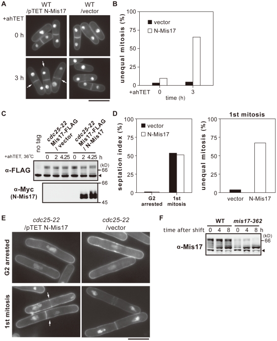
Figure 3. Rapid occurrence of missegregation by N-Mis17 under the TET promoter.
A. Chromosome missegregation was frequently observed after TET-induced overproduction of N-Mis17 in wild-type cells at 33°C as shown in the DAPI-stained micrograph (arrows). Bar, 10 µm. B. N-Mis17 was expressed under the TET promoter. The frequencies of chromosome segregation (%) were measured at 0 and 3 h after induction by the anhydrotetracycline (ahTET) at 33°C. C–E. The cdc25-22 mutant was transformed by plasmid carrying N-Mis17-Myc expressed under the TET promoter. Endogenous Mis17-FLAG was chromosomally integrated and expressed under the native promoter. Mis17-FLAG and N-Mis17-Myc were assayed by immunoblotting using antibodies against FLAG and Myc. Arrowhead indicates the cross-reacting antigens. The cdc25-22 mutant cells that expressed endogenous Mis17-FLAG and ectopically produced N-Mis17-Myc were arrested after 4.25 h at 36°C. See text for explanation of (C). After the release of mutant cells to 26°C, the frequencies of chromosome missegregation and the septation index were measured (D). Micrograph of missegregation (arrows) is shown in (E, Bar, 10 µm). F. Immunoblotting was done to detect Mis17 protein in the extracts of the wild-type and ts mutant mis17-362 cells at 36°C for 0–8 h. The mis17-362 mutant protein was unstable. Arrowhead indicates the cross-reacting antigens.

