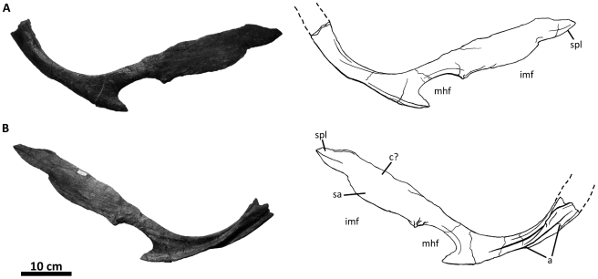
Figure 30. Left prearticular of Acrocanthosaurus atokensis (NCSM 14345).
Prearticular in (A) medial and (B) internal views. Dashed lines represent material not in figure. a, angular contact; c, coronoid contact; imf, internal mandibular fenestra; mhf, mylohyoid foramen; sa, surangular contact; spl, splenial contact.

