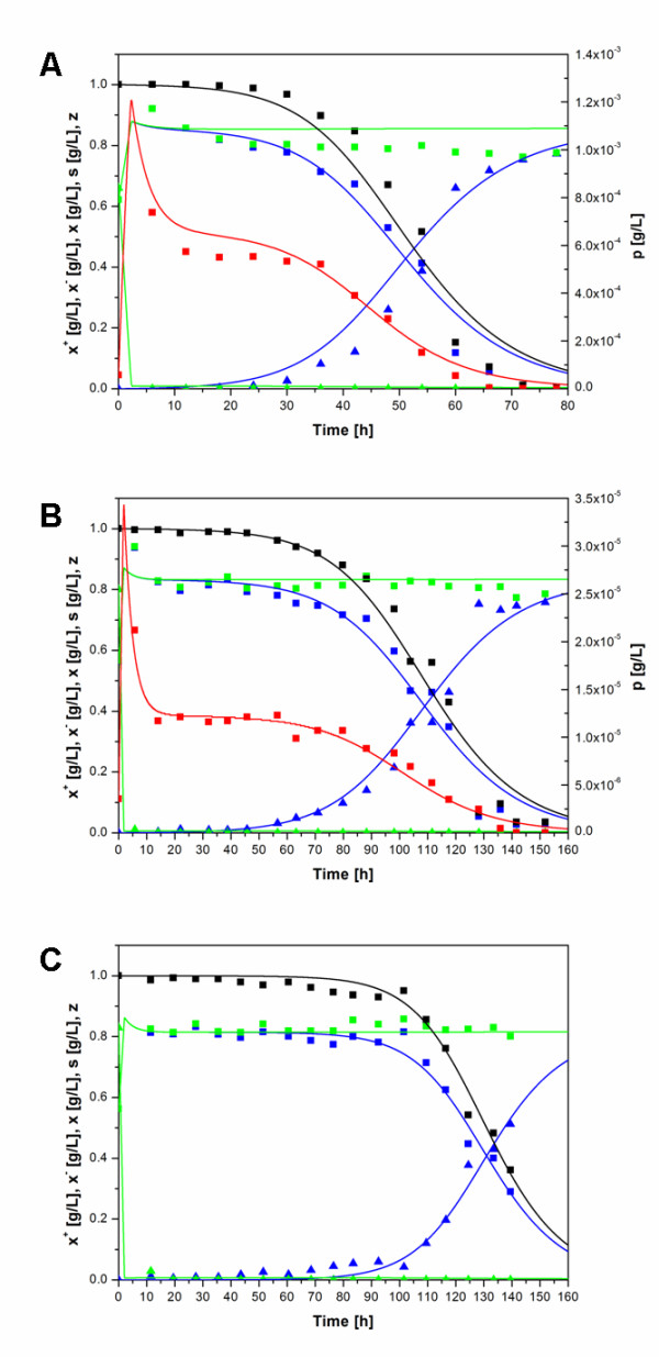
Figure 3.

Growth and plasmid loss kinetics of transformed E. coli LE392 cells in a chemostat culture. Experimental data (data points) and simulation curves obtained by the model of Lee et al. (lines): plasmid-harbouring biomass, x+ (blue squares and line); plasmid-free biomass, x- (blue triangles and line); total biomass, x (green squares and line); glucose concentration, s (green triangles and line); hIFNγ concentration, p (red squares and line) and plasmid-harbouring cell fraction, z (black squares and line) for cells carrying the plasmid pP1-(SD)-hIFNγ (Figure 3A), pP1-(4SD)-hIFNγ (Figure 3B) and pP1-(ΔSD)-hIFNγ (Figure 3C).
