
Figure 1.
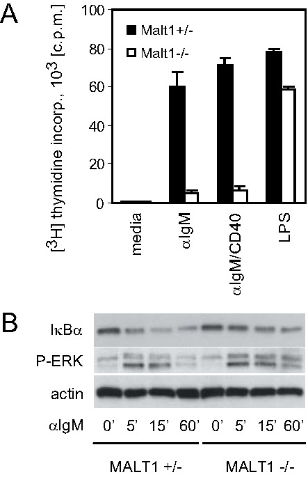
Activation of BCR signaling in MALT1-/- B cells. (A) B cell proliferation. Splenic MALT1+/- and MALT1-/- B cells were stimulated for 36 h with soluble anti-IgM, anti-IgM plus anti-CD40, or LPS. Proliferation was measured by [3H]thymidine incorporation. Results are presented as the mean [3H]thymidine incorporation ± S.D. for triplicate samples after an 8 h pulse and are 1 trial representative of at least 3 independent experiments. (B) IκBα degradation. Splenic MALT1+/- and MALT1-/- B cells were stimulated for the indicated times with anti-IgM. IκBα degradation, ERK1/2 phosphorylation (P-ERK), and actin levels were determined by Western blotting.