
Figure 4.
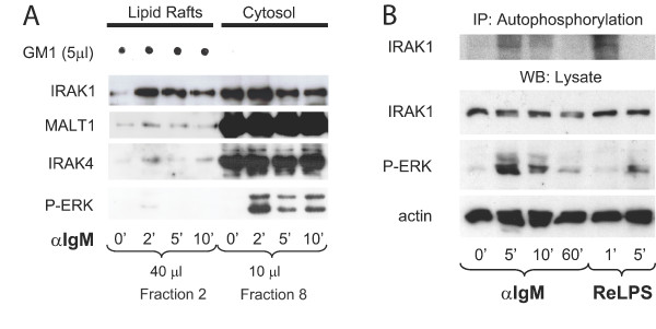
Lipid raft recruitment and activation of IRAK1 and IRAK4. (A) Lipid raft recruitment of IRAK1 and IRAK4. Splenic WT B cells were left unstimulated or were stimulated for the indicated times with anti-IgM. Lysates were separated by sucrose-gradient ultracentrifugation and the indicated amounts of the collected fractions were analyzed by Western blotting with antibodies to IRAK1, MALT1, IRAK4, and phospho-ERK. To identify the lipid raft containing samples, 5 μl of each fraction were spotted onto a nitrocellulose membrane and GM1 was visualized with FITC-labeled Cholera toxin. (B) Activation of IRAK1 upon BCR triggering. Splenic WT B cells were stimulated for the indicated times with anti-IgM or ReLPS. The kinase activity of immunoprecipitated IRAK1 was measured by an autophosphorylation assay. IRAK1, phospho-ERK1/2, and actin were quantified by Western blotting of the lysates. IP, immunoprecipitation; WB, Western blotting.