Figure 5.

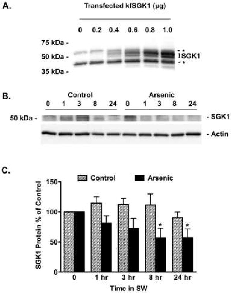
Arsenic reduced the abundance of SGK1 protein in the gill of fish transferred from freshwater (FW) to seawater (SW). A) HEK cells were transfected with kfSGK1 (0, 0.2, 0.4, 0.6, 0.8 or 1 μg cDNA) and SGK1 abundance was measured by Western blot. The nonspecific protein bands, indicated by the dashed lines and asterisks, are most evident in the lanes marked 0 and 0.2 μg cDNA. As shown previously, SGK1 is a double ∼50 kDa (Shaw et al., 2008), and its abundance increases as a function of the cDNA concentration. By contrast, the nonspecific bands do not change. This experiment demonstrates that the SGK1 antibody from Sigma recognizes kfSGK1. Fish were adapted to freshwater (FW) for two weeks, then exposed to arsenic (12,000 ppb) or vehicle for 48 hours and then placed in seawater (SW) containing arsenic (12,000 ppb) or vehicle for 1 hr, 3 hr, 8 hr and 24 hrs. B) Representative Western blots of kfSGK1. C) After the indicated times, gills were isolated and SGK1 protein in control (hatched bars) and arsenic treated fish (solid bars) were measured as described in methods. Data presented as percent of time 0. N=7/group. *P<0.05 versus control.
