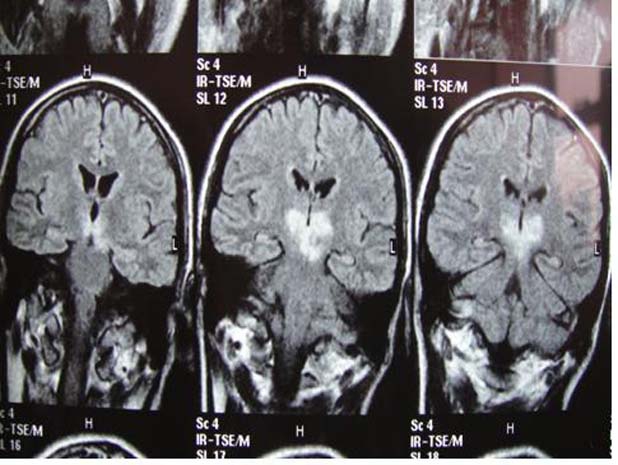
Figure 3.

Coronal T2 FLAIR brain MRI of our first patient. Note the persistent hyperintense signal abnormality (which did not suppress on the FLAIR film) at the paramedian thalami and the left side of the rostral midbrain. The patient presented with acute thalamopeduncular syndrome.
