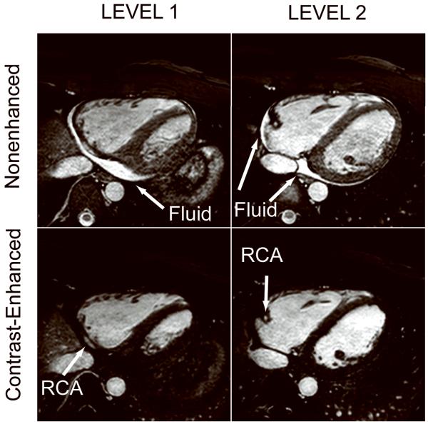
Figure 4.
Example of nonenhanced (top row) and contrast-enhanced (bottom row) whole-heart SSFP coronary images acquired in a subject. The epicardial fluid has high signal intensity on the nonenhanced SSFP coronary MRI, which may compromise depiction of adjacent middle and distal RCA. The fluid is completely suppressed on the contrast-enhanced MR images.

