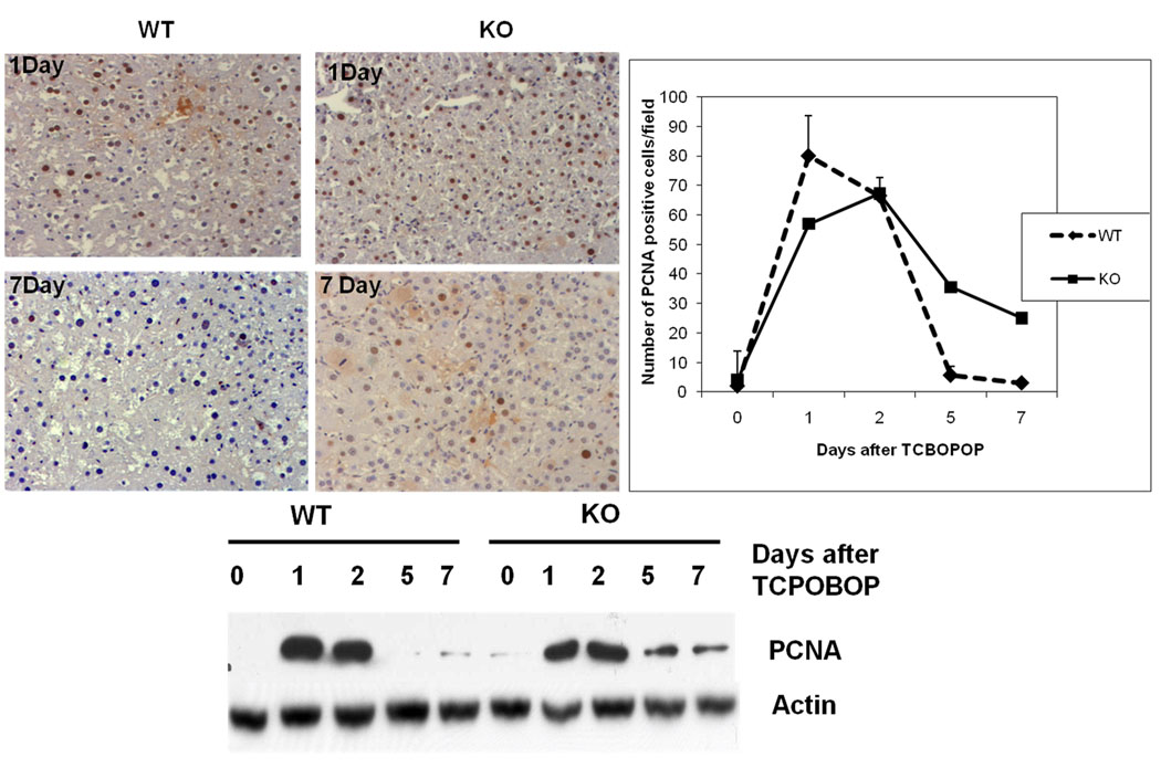
Fig 2.
Cell proliferation kinetics after TCPOBOP administration (A) Hepatocyte proliferation was quantitated by counting the PCNA positive cells. PCNA positive cells were counted in low-power fields (200×) in 3 sections from 3 different ILK/Liver −/− or WT livers. Each data point is the mean ± SE from more than three measurements per point. (B) Representative photomicrographs of PCNA stained liver section of ILK/Liver−/− and WT mice at day 1, and 7 after TCPOBOP administration. (C) Western Blot analysis of PCNA. Pooled liver samples from at least 3 mice were used for protein analysis.

