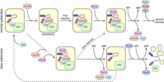
FIGURE 1.
Schematic of proteins joining and leaving the spliceosome surrounding the actions of Prp16. Two pathways, one for normal (wild-type) substrates (upper) and one for slow (mutant) substrates (lower), are shown. Steps that occur relatively quickly are shown with unbroken arrows, whereas those that occur relatively slowly are shown with dashed arrows. The spliceosome is shown in yellow; exon 1 as a blue rectangle; the intron as a purple line; exon 2 as a magenta rectangle; the NTC (Nineteen Complex) in green; Yju2 in light blue; Cwc25 in orange; Prp16 in pink; Prp43 in blue; Prp22 in green; Prp18 in red; and Slu7 in purple. ATP hydrolysis-requiring steps are indicated. Yju2 can, but does not necessarily, join the spliceosome early, but it must join before Cwc25. Cwc25 may join as shown here, but the spliceosomal species labeled “transient” has not been detected; Cwc25 may join after the first step, but it must join before Prp16. For slow substrates, the overall pathway is likely slower than for normal substrates (as indicated by the first dashed arrow), and the “first reaction” for slow substrates must be slower than the Prp16-rejection pathway.

