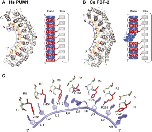
FIGURE 1.
PUF protein–RNA interaction. (A) Hs PUM1: structure of human Pumilio1–RNA complex (PDB accession code 1M8Y) (Wang et al. 2002). (B) Ce FBF-2: structure of C. elegans FBF-2–RNA complex (PDB accession code 3K5Y) (Wang et al. 2009b). Eight RNA-recognition helices (gray in structures and diagrams) contribute side chains to contact RNA bases (blue). (Red) Stacking residues; interactions with base edges are not shown. (Beige shading) Amino acid side chains and RNA bases involved in stacking interactions. (C) Key interactions in the FBF-2–RNA complex. (Red) Stacking residues; (green) base edge interacting residues; (blue) RNA bases.

