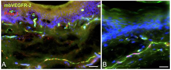
Figure 1.
Immunostaining of membrane-bound VEGFR-2 (mbVEGFR-2) in human foreskin (green) shows its localization on blood vascular and lymphatic endothelial cells (the latter are red by anti-Prox-1 nuclear staining). Dapi staining (blue) was used to demonstrate cell nuclei. A) Bar = 100μm. B) Bar = 50μm.

