
Figure 3.
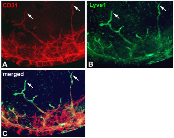
Immunostaining of the cornea of a LeCre;svegfr-2loxP/loxP newborn mouse, which selectively lacks svegfr-2 in the cornea. A) CD31, a pan-endothelial cell marker is stained red corneal vessels. B) A specific marker for lymphatic endothelial cells, Lyve-1 (lymphatic vascular endothelial hyaluronan receptor-1) stains corneal lymphatics in green. C) Merged image of CD31 and Lyve-1 staining demonstrates that all vessels within the cornea are lymphatic vessels, whereas in the limbus both types of vessels are present. The arrows point to typical blind-ended morphology of lymphatic vessels.