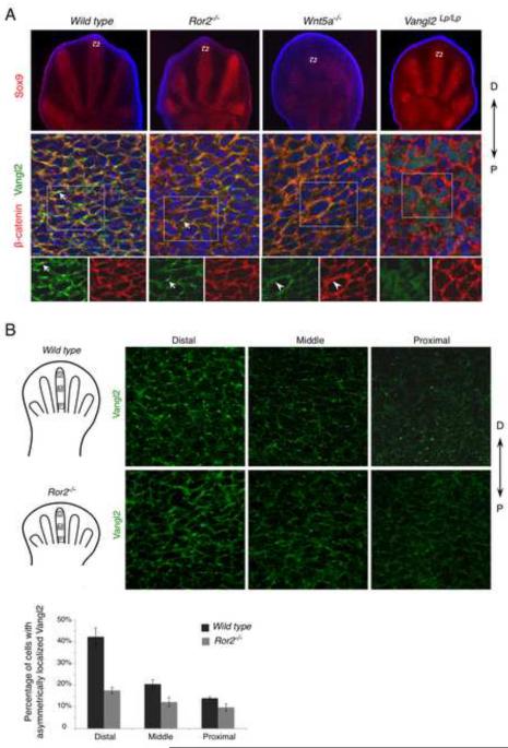
Figure 4. Wnt5a is required for Vangl2 asymmetrical localization.
(A) Digit chondrocytes of E12.5 embryos is shown by Sox9 expression (red) (upper panel). The selected regions (boxed) are enlarged and shown with Vangl2 (green) and β-catenin (red) co-staining (projected Z-stacks) (middle panel). The boxed regions are shown in lower panel with separate staining of Vangl2 and β-catenin. Vangl2 asymmetrical localization (arrow) is completely lost in the Wnt5a−/− limb. In the Ror2−/− mutant limb, Vangl2 asymmetrical localization was lost in the more mature cartilage (see Fig. S4A), but still detectable in the distal limb bud with reduced intensity (arrow). Vangl2 is still localized to the cell membrane in Wnt5a−/− mutants (arrowhead). D, distal; P, proximal. (B) Projected Z-stack pictures of Vangl2 staining (green) from distal (D), middle (M) or proximal (P) parts of E12.5 wild type or Ror2−/− embryonic distal limbs (schematized in left panel). The number of cells with Vangl2 asymmetrical localization is progressively reduced from distal to proximal parts and much reduced in the Ror2−/− mutant. Quantified results were shown in lower panel. Error bars are ± SD, n=2. See also Figure S4.

