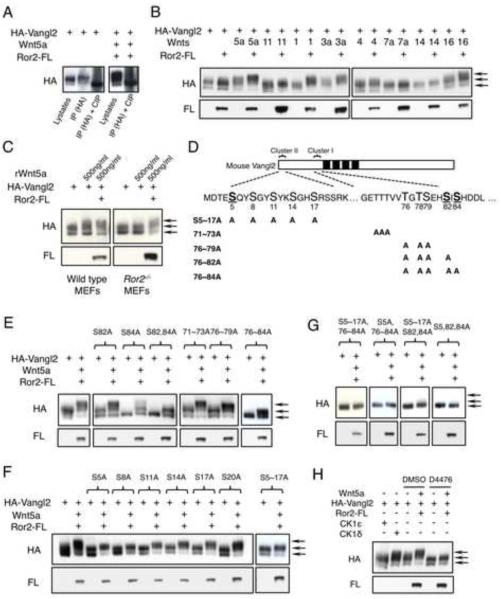
Figure 5. Wnts act through Ror2 to induce Vangl2 phosphorylation.
(A) Mobility shift of Vangl2 expressed in CHO cells on SDS-PAGE gel was enhanced by Wnt5a, but abolished after treatment with calf intestine phosphatase (CIP). (B) CHO cells were transfected to express different mouse Wnts, Ror2 and Vangl2 in the indicated combination. Vangl2 phosphorylation was analyzed by immunoblot. Arrows indicate hyperphosphorylated (upper), hypophosphorylated (middle) and unphosphorylated (lower) Vangl2 forms. (C) MEF cells were treated with recombinant Wnt5a protein for 2 hours and Vangl2 phosphorylation was analyzed. (D) Mouse Vangl2 with two phosphorylation clusters. Founder sites of Cluster I and II are underlined and shown in bold. Mutant Vangl2 with various serine/threonine (S/T) to alanine (A) variants are shown. (E) S84 and S82 are founder sites for Cluster I phosphorylation. Phosphorylation of Cluster II sites still occurred when all Cluster I sites were mutated. (F) S sites in Cluster II of Vangl2 were individually mutated to A. Wnt5a-induced Vangl2 phosphorylation was progressively more reduced from S20A to S5A. (G) Mutating all three founder sites, S5, 82, 84, is sufficient to completely abolish both basal level and Wnt5a-induced phosphorylation in CHO cells. (H) Vangl2 phosphorylation is induced by CK1δ not CK1ε and partially blocked by CKI inhibitor D4476 (100uM) in CHO cells. See also Figure S5.

