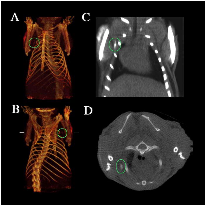
Figure 5.

CT imaging of a lymph node of a mouse with a bismuth nanoparticle (a,b) Three-dimensional volume renderings of the CT data set, the length of the reconstruction is 3.8 cm. (c) Coronal slice (length of the slice 2.3 cm). (d) Transverse slice at the height indicated by the horizontal lines in b. The maximal diameter of the mouse is 1.8 cm. The position of the lymph node under the right shoulder is indicated by the ovals, and the arrows show the injection site. Note the lack of contrast in the corresponding contralateral (left shoulder) lymph node. Reprinted with permission from [50].
