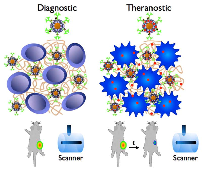
Figure 6.

Schematic drawing of the theranostic approach. The diagnostic particle consists of a diagnostic moiety and a coating, modified with a nuclide (green), targeting an antigen (brown), here deposited between the tumor cells. Imaging is performed with MRI and PET. The particle can carry a payload drug (red) that is released in the tumor creating a theranostic agent, allowing for imaging and therapy of the tumor.
