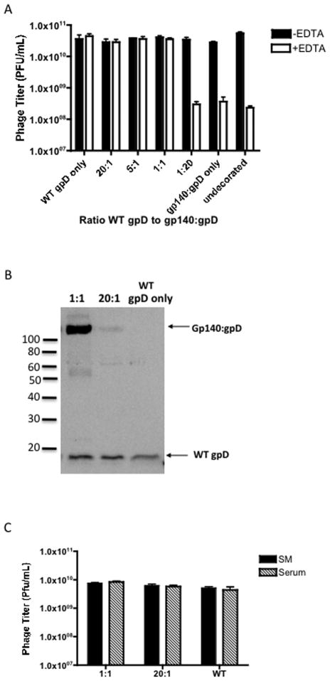
Figure 4. Stability of Env decorated bacteriophage particles.
(A) 1×10⁹ PFU (plaque-forming units) of gpD-deficient bacteriophage lambda procapsid were decorated with varying ratios of WT gpD protein and gp140:gpD fusion protein. The decorations were performed by incubating the desired molar ratios of proteins with the gpD-deficient phage (1× 10⁹ PFU) at 30°C for 20 minutes. The stability of the decorated phage samples was then tested by the addition of 100mM EDTA (see Fig. 2) and titering on LE392 bacteria. Undecorated phage was analyzed as a control (EDTA sensitive). The decorations were performed in triplicate within the same assay. (B) 1L preparations of phage decorated with 1:1 or 20:1 WT gpD to gp140:gpD, or WT gpD only were prepared by CsCl-banding and dialysis, titered on LE392 E.coli host cells and then loaded on a 12.5% SDS-PAGE gel, (109 PFU/lane). Phage protein content was examined by immunoblot analysis, using antiserum directed against gpD. Incorporation of both wild-type gpD and gp140:gpD fusion protein in the phage preparations decorated with 1:1 or 20:1 WT gpD to gp140:gpD is indicated by the arrows. (C) 1:1, 20:1 and wild-type (WT) phage (1 × 10¹⁰ PFU/ml) were incubated for 30 min at room temperature in SM buffer or normal rabbit serum and subsequently titered on LE392 bacteria to determine stability. The data are represented as means ± SEM.

