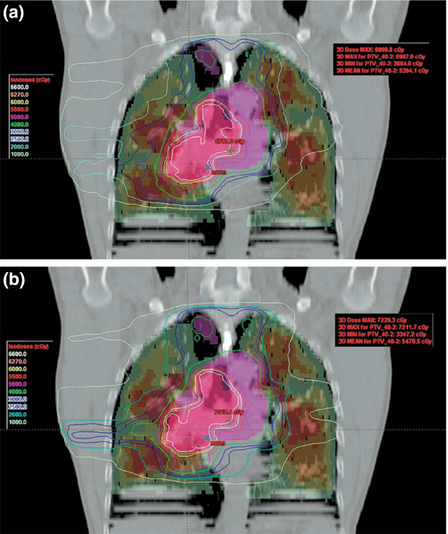
Figure 1.
Coronal view showing plans with (bottom) and without (top) SPECT guidance. The central purple and pink structures are primary and boost targets; lung is shaded by SPECT activity intensity, ranging from red (highest) to green (lowest). Isodose lines demonstrate that SPECT-guidance decreased dose to higher perfusion regions. Reprinted with permission.30

