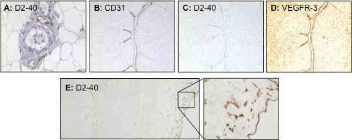
Figure 2.
Immunostaining of D2-40 in systemic vessels, and D2-40, CD31, and VEGFR-3 in placental umbilical cord. A: D2-40 staining in maternal vessels from normal pregnant women. Positive D2-40 staining is only seen in lymphatic vessel endothelium, but not in arterial or venous blood vessel endothelium. B, C, and D: Immunostaining of CD31, D2-40, and VEGFR-3 in umbilical cord, respectively. Negative D2-40 (C), but positive CD31 (B) and VEGFR-3 (C), staining is seen in umbilical vein endothelium. E: Positive D2-40 staining is also observed in cord membrane (amniotic epithelium) and underlying matrix (Wharton's Jelly) (E: insert).

