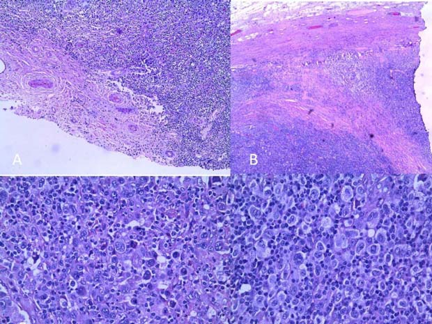
Figure 1.

(A) Diffuse destruction of testicular tissue sparing only part of capsule with adventitial vessels. (B) Broad collagenised fibrous bands. (C) Rare diagnostic Reed Sternberg cell and a few mummified cells. (D) Cellular area showing numerous mononuclear and lacunar atypical RS cells, with a scanty background lymphocyte population.
