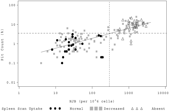
Figure 1.
HJB versus PIT counts. Individual patients are represented by a symbol on the scatter plot corresponding to their qualitative LS scan result. (●●●) represents patients with normal splenic uptake; (▩▩▩), patients with splenic function that is present but decreased; and (▵▵▵), patients with absent splenic function. The dashed horizontal line denotes a log10 PIT count of 3.5%; and the vertical dashed line, log10 HJB of 300/106 RBCs.

