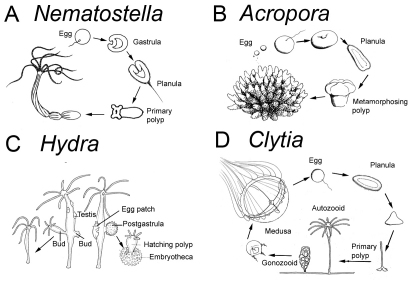
Fig. 2.
Life cycles of the main cnidarian model systems. (A,B) Anthozoan polyps either burrow into the soft substrate (A), here exemplified by the edwardsiid sea anemone Nematostella vectensis, or are attached to the surface (B), as with many sea anemones and corals, here exemplified by Acropora millepora. Nematostella female polyps release packages of several hundred eggs into the water, where they are fertilized (Fritzenwanker and Technau, 2002; Hand and Uhlinger, 1992). The resulting embryos develop into ciliated planula larvae that undergo either a gradual (Nematostella) or more dramatic (Acropora) metamorphosis into a sessile primary polyp, which involves calcification and formation of the skeleton in corals. (C) In Hydra, gametes develop from interstitial stem cells located in the ectoderm that differentiate within several testes or within a single egg patch. The embryo remains attached to the parent polyp from fertilization through gastrulation. The postgastrula embryo forms a cuticle from which the primary polyp hatches after several weeks or months. (D) The hydrozoan Clytia hemisphaerica forms a colony with feeding polyps (autozooids) and medusae-bearing gonozooids. Gametes are released from the medusae into the water. The embryo develops into a planula larva that settles to transform into a primary polyp, which then forms a new colony. Drawings are by Hanna Kraus (A,B,D) or modified from Tardent (Tardent, 1978) with permission (C).

