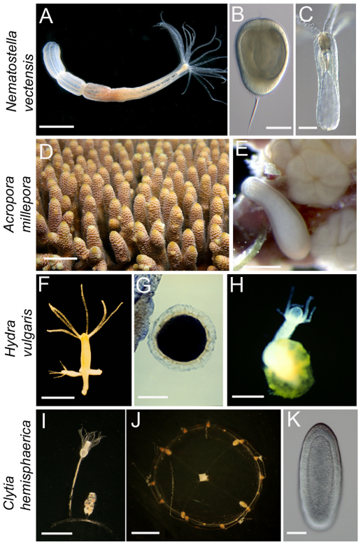
Fig. 3.
Cnidarian model systems used in developmental biology. (A-C) Nematostella vectensis, showing adult polyp (A), planula larva (B) and primary polyp (C). (D,E) Acropora millepora showing coral (D) and planula larva and metamorphosing early settlement stages (E). (F-H) Hydra vulgaris showing budding polyp (F), cuticle stage postgastrula embryo (G) and hatching primary polyp (H). (I-K) Clytia hemisphaerica showing autozooid and gonozooid polyps (I), young medusa (J) and planula larva (K). Note the differences in size between different cnidarians. All polyps and planulae are oriented with oral side up (except for A). Images were taken by Jens Fritzenwanker and U.T. (A-C), David Miller (D), Eldon Ball (E), Tim Nüchter and Thomas Holstein (F), U.T. (G,H) and Hanna Kraus and U.T. (I-K). The images in B and C are reproduced with permission (Rentzsch et al., 2008). Scale bars: 1 cm in A; 70 μm in B,K; 80 μm in C; 5 cm in D; 150 μm in E; 500 μm in F,J; 250 μm in G,H; 100 μm in I.

