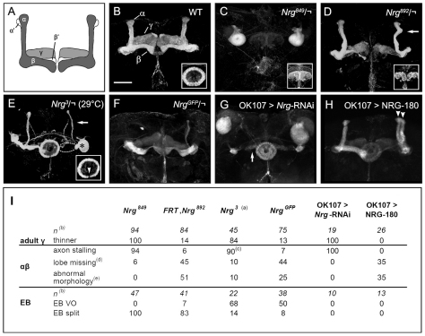
Fig. 1.
Neuroglian mutant phenotypes. Whole-mount anti-FAS2 staining of adult central brain labels EB and α, β and γ MB lobes (see B). All images are projections of confocal z-stacks. Scale bar: 50 μm. (A) Schematic representation of Drosophila MBs with α, α′, β, β′ and γ lobes. (B) Wild-type MB lobes and EB (inset). (C) MB axon stalling and a split EB (inset) in hemizygous Nrg849. Only the axon stalling and EB defects are shown in this confocal stack. Gamma lobe phenotypes are the same as in G (see below). (D) Nrg892 hemizygote with β lobe missing MB phenotype, thinner γ lobes, abnormal α-lobe morphology (arrow) and split EB phenotype (inset). (E) MB and EB defects in hemizygous Nrg3 grown at restrictive temperature (29°C). MB phenotypes include thinner (arrow) or missing (dashed line) lobes and axon stalling (asterisk). The EB has a ventral open phenotype (arrowhead). (F) NrgGFP hemizygote with thin or missing α lobes, β lobes with outgrowth defects and thinner γ lobes. (G) MBs from OK107>Nrg-RNAi with axon stalling defects, and a prominent γ lobe defect with thin, often abnormally oriented, lobes (arrow). The EB is wild type, as OK107-GAL4 does not drive expression in this neuropil. (H) Lobes of NRG-180 overexpressing MBs show guidance defects (right brain hemisphere): the majority of β axons project vertically, parallel to the α lobe, resulting in two vertical lobes (arrowheads). (I) Mushroom body phenotype summary. (a) At 29°C from first instar larval stage until dissection as pharate adults. (b) n for MB phenotypes is the number of brain hemispheres analyzed, while n for EB phenotypes is the number of brains analyzed. (c) Complete (all axons) or partial (majority of axons) axon stalling. (d) Lobe missing caused by axon branching and/or axon guidance defects. (e) The defects in MB lobe morphology (referred to as abnormal morphology) for the different genotypes is described in detail in the text.

