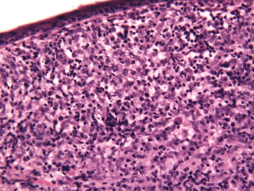
Figure 2.

Histologic characteristics: The epidermis is thinned. Within the dermis, there is a nodular infiltrate of lymphocytes and enormous numbers of macrophages touching the epidermal junctional zone and reaching down to the superficial subcutis. The cytoplasm of macrophages (mainly within the papillary dermis) is filled with numerous Leishmania amastigotes. (Hematoxylin and eosin stained original magnification ×40).
