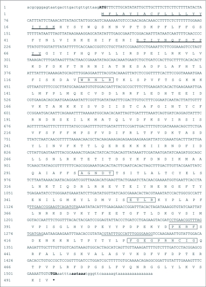
Figure 1.
Nucleotide and deduced amino acid sequence of CYP345D3. The nucleotides underlined show the positions of gene specific primers used in the experiment. The start codon ATG is indicated with bold and the stop codon TGA is indicated with bold and by an asterisk. Polydenylation signal AATAAA is shown in bold italic lowercase letters. The heme-binding sequence motif FxxGxxxCxG and other sequence motif are indicated by the boxed amino acids. The transmembrane domains are shown in deeply underlined amino acid residues.

